+7(499)344-03-03
Register for reception Online consultation
The whole site
Register for reception Online consultation
Oganes E. Dilanyan
Urologist, MD, PhD

Ureteral stones

Reading time: 4 min.
In this article

Ureter stones is the worst manifestation of urinary tract stones.

The ureter is a very thin tube 3mm in diameter which connects the kidney with the bladder and any stone travelling from the kidney  acts as a plug and disrupts urinary flow. The urine collects in the kidney stretching it. This causes sharp pain. 

Ureteral stones symptoms

Specific features: Almost always a sharp pain in the kidney or kidney colic. In the vast majority of cases infection can develop and this sometimes can lead to losing a kidney. In some cases, it can be quite difficult to treat an ingrown stone with Ureter widening. It would seem a small stone can cause quite big problems.

Removing the stone from the ureter – strange as it may seem this is not a priority for the doctor. The first priority is to restore urine outflow as stagnating urine is the perfect breeding ground for bacteria which can lead to purulent pyelonephritis, quite a serious condition. One has to agree that it would be silly to lose one’s kidney or possibly even one’s life because of one tiny stone. 

Ureteral stone: surgery

image/svg+xml

You wish. Do you think we are going to operate straight away? Not a chance. As I said, first of all we need to restore urine outflow.
Ureter stenting – the procedure is used to place a stent which is a hollow tube into the ureter containing the stone. The stent allows to restore urine flow.
Breaking up the stones – in more than 50% of cases it is possible to carry extracorporeal shockwave lithotripsy and often without the stenting.
Removal of the stone from the ureter endoscopically without any incisions or using laser lithotripsy in those cases where the stone cannot be broken down or passed out. 
 

What to do if the stone is stuck in the ureter

Pain. Coming in waves, heavy and exhausting pain is almost always the hallmark of the ureter stone. Simply because even a 3-4 mm stone can block urinary flow.  The urine collects in the renal pelvis, the pelvis widens and tries to contact resulting in a spasm that some women often compare to labour pains.  The affected person wants only one thing – to get rid of the pain any way possible be it tablets or injections, just for the pain to go away. 
Often the person can feel sick, the blood pressure may go up and if the stone is positioned in the lower part of the ureter close to the bladder frequent urge to urinate also occurs even though the bladder is empty.

As humans we understand that no one wants to experience this sort of pain. The timing is always bad, it always disrupts one’s plans be it a business meeting, a business trip or even a holiday. But it is very important to know that killing the pain is not going to cure it. It is possible to suppress the pain for a few days and then the kidney will simply stop hurting. One may think great! The pain is gone! But there are two hidden dangers here. Firstly, if infection develops in the ureter it can lead to purulent pyelonephritis and sepsis which often leads to kidney loss. Secondly, the kidney can just die off slowly and turn into a useless bag containing some urine and a stone.

“Rockfall is the Earth’s terrible and uncontrolled weapon”
Sun Tzu “The Art of War”

Where do these ureteral stones come from?

Almost in all cases the stone gets into the ureter from the kidney. It can either be a small stone or a chip off a bigger stone. Kidney stone is a rather useless thing. It would seem it sits in the kidney and grows slowly without causing any pain. And then…. A bumpy ride, a run, or even sometimes vigorous intercourse can force the stone downwards from the kidney. The stone in this case acts in the same was as a plug would inside a hose – it disrupts the flow. Urine stagnation begins, the renal pelvis widens and here we have the episode of acute kidney colic. There are several possible scenarios:

•    The stone is small and will make its own way out. One can be hopeful if the stone is not big and the patient does not suffer from ureter narrowing. In any case at least a kidney ultrasound should be performed.
•    Developing an infection can lead to acute pyelonephritis. This is quite serious and requires emergency measures such as kidney stenting to restore urine outflow as well as an antibacterial treatment. 
•    The patient will take pain killers for several days until the kidney “goes quiet”, in other words it will stop producing urine. The pain will go away but in a few months’ time it will be discovered that the kidney had died and turned into a useless urine containing bag which needs to be removed.

Ureteral stones in both men and women are not only the cause of severe pain but also carry a real threat of purulent pyelonephritis and kidney loss.

From personal experience

… “A 34-year-old patient with kidney stone disease presented at the clinic at 3 am with a stone in the lower third of the right ureter and symptoms of a kidney colic”. – reports the doctor on duty during the morning meeting. “We have done all the necessary tests and investigations urgently and the patient is pain free and has slept the rest of the night”. 
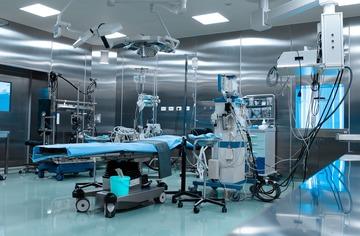
We examine the CT scan of the kidney. The stone is almost one cm in size. Usually those get stuck just as they are leaving the kidney. The stone density is over 1000 Hounsfield units which makes extracorporeal shockwave lithotipsy impossible.    
- Tell me please where is the pain and when did it start? – I ask the patient. 
- I knew I had a kidney stone, - says the patient with great annoyance in his voice. But I kept putting it off. Yesterday we signed a contract, a had a little bit to drink, got a taxi home. I live outside Moscow, the road is quite bad, it was a bumpy ride. It started as soon as I got home. Back pain radiating into the groin, almost unbearable. It was hurting the same standing up and lying down. Thanks to the doctor, everything got sorted out quickly, he did an ultrasound and injected a painkiller. I am flying tomorrow evening. I cannot miss the trip as the contract is riding on it.  
- Well, you did not have a temperature and there is no kidney inflammation. I think we can operate and send you on your flight tomorrow – I smile at him, - Please do not eat or drink anything, we will start in about an hour and a half. 
- What do you mean operate? – The patient is terrified. – I will be in bed for 2 weeks after the operation. 
-Well… Endoscopy is also an operation. Don’t worry. We will break the stone up with a laser and you will be good as new tomorrow following the operation. We will insert a stent just in case just to safeguard you from any mishaps during your flight. I draw a diagram of the procedure and explain why he can expect quick recovery. Just don’t eat anything, ok?

A tiny spark of fire has a power to burn down a whole city

Ureteral stones diagnosis

There is a number of tests that need to be performed quickly in the presence of stones.   

Ureteral stones diagnosis

General Urine test

Is done straight away due to extremely high risk of purulent pyelonephritis and this test helps determine the severity of the inflammation.  

Full blood count

Is also performed as soon as possible for the same reason. The risk of purulent pyelonephritis affects the number of white blood cells and reflects the body’s inflammatory response. 

Biochemical blood test

This test consists of a number of parameters and is strictly necessary in order to understand the condition of the body. For example, creatine levels can warn us about impaired kidney function and glucose will alert us to diabetes. 

Kidneys ultrasound

This allows us to assess the widening of the kidney, the rate to which the blood supply to the kidney is interrupted and, in some cases, will allow us to see and assess the size of the kidney stone. This is the first visual visualising test in the presence of this condition. 

Intravenous urography

This is a radiological method which helps to determine several aspects such as ureter stone size, it’s position, the degree of kidney function impairment and the presence of other stones. 

CT scan

We often replace urography with a CT scan which allows us to gain the maximum amount of information including information about stone density. This determines the effectiveness of the treatment.

Ureter stones are the main reason why good urologists lose their sleep

Ureteral stone removal surgery

In the original version of the Hippocratic one can see the following words:” I will not use the knife, even on sufferers from stone”. It is rather strange but in modern urology these words are once again very relevant. In a well-equipped clinic the number of operations involving cutting up of a patient with kidney stone disease should be kept to a minimum.  There are several methods to break down the stones and remove the stones from the ureter without surgery or use of a scalpel.

Methods of renal duct stone removal surgery

Extracorporeal shockwave lithotripsy

Basically, this procedure allows us to break down ureter duct stones in both men and women without any surgery. An ultrasound wave passes through the stone breaking it into small pieces which then pass out of the body on their own accord. Unfortunately, not all stones can be broken down in this manner. The denser the stone the less success ultrasound will have in breaking it down.

Laser lithotripsy

This method involves placing a laser fibre next to the stone and the stone breaks up into dust. The particular. The laser fibre needs to somehow get to the stone. For this we use both hard and soft endoscopes which allow us to get to the stone through the urinary tract without making a cut.

Laparoscopic removal of the stone

In rare cases, for example in the presence of ureter narrowing or if the stone has been in the ureter for a very long time it is simply impossible to remove it endoscopically or break it down. In the past these cases warranted abdominal surgery but now even these stones can be removed laparoscopically using 3-4 punctures instead of one ugly cut.

Laser Lithotripsy

Having modern equipment in our clinic allows us to perform laser lithotripsy without any need for an incision. A thin endoscope is passed through the urethra into the ureter and we are able to see the stone on the screen. We approach it with the laser which breaks the stone into dust. The patient feels no pain following this intervention and can be discharged home the following day in the majority of cases. The video bellow shows how this procedure is performed.  

From personal experience
- Are you ready? May we, Andrey? Let’s roll and may God be with us…. This is a traditional start to all surgical procedures. First, we check the team is ready, then ask for the anaesthetist’s permission to start and God’s help never goes amiss. - Right. Pass the ureteroscope please. Theatre nurse passes a long and thin endoscope which I use to get into the bladder via the urethra. I look around. Everything is fine here, moving on! I get into the ureter reaching the stone. Here it is, a boulder! And look at the swelling! Pass the laser. I move the laser fibre to the stone and start breaking it up, turning it to dust, as mentioned before. - Please gather some of this to be sent for a chemical analysis – I say to my team even though they always do it on autopilot anyway. – And pass the stent please. The stone is broken down. But due to it being in there for so long the mucosa is very swollen. The swelling could block the opening and the patent would be in pain once again. Hence the stent. -That’s it, we are finished. Thank you everyone! – another traditional phrase for it is not only me who has done all the work, it was a team effort. The following day. - How are you feeling? – I smile at him anticipating his reply. The kidney ultrasound concluded the kidney was not widened. Are you ready for your flight? - Rearing to go! – reports the patient, - Slight burning during urination but it does not bother me. - Well, all the best. When you come back in a week we will remove the stent. And the stone analysis will be back by then. - What sort of analysis? – The patient looks suspicious. Should I be expecting something bad? - No, - I smile. We need to understand where the stone came from. We will prescribe a special diet so the situation does not repeat itself. - Oh yes, no repeats please. A shadow of suffering briefly touches my now ex-patient’s face. You cannot even imagine the pain …
Our Advantages

Professional Approach: modern diagnostic methods of the causes and complications of ureter stones. 

Our team: Doctors specialising in endoscopic urology.

Equipment: endoscopic urology operating theatre equipped with Karl Storz and Olympus equipment.

Board of doctors: For your safety we have the following doctors available: a urologist, a cardiologist and an anaesthetist.

Safety: we only use methods recommended by professional associations such as EAU and AAU.

Comfort: no queueing or waiting for a consultation and premium inpatient facility.

Result: removal of ureter stones and their causes. 

We are also GCP certified, which confirms we provide the highest standards of diagnosis and treatment. 

Certificates
image/svg+xml
image/svg+xml
Our Guarantees

-We guarantee your safety by using modern and highest quality anaesthesia manufactured in Austria or France. 
-We DO NOT USE low quality equivalents of any drug which significantly lowers the risk of any complications
- Using a modern endoscope by Karl Storz guarantees successful laxer treatment to all my patients. 
- Working in the field of endoscopic urology in Austria and Italy and extensive experience in treating kidney stone disease ensure the absence of complications.   
-We DO NOT REFER our patients to unknown clinics for follow up and further treatment and provide outpatient follow up as well as inpatient treatment.

Every patient is provided with a discharge summary with detailed recommendations and an official sick leave certificate
Information for foreign patients:

If you require ureteral stones surgery in Moscow please contact us. We will arrange admission to hospital on the same day you arrive and will carry out all the necessary tests and investigations and perform the surgical procedure as soon as possible. You will be able to return home after 3-4 days

image/svg+xml
image/svg+xml